
The GLP-2 receptor was isolated by expression cloning using an initial strategy employing degenerate PCR primers to amplify novel glucagon/GLP-1 related receptors Prototypic G protein-coupled receptor for the intestinotrophic factor glucagon-like peptide 2 Proc Natl Acad Sci U S A. 1999 Feb 16;96(4):1569-73. The GLP-2 receptor exhibits ~ 50 % amino acid identity with the GLP-1 receptor, and clearly represents a new member of the glucagon-secretin receptor superfamily. The human GLP-2R has been localized to human chromosome 17p13.3 by FISH. No GLP-2R linkage to specific human diseases has yet been reported.
The cloned GLP-2 receptor responds to GLP-2 via activation of adenylate cyclase and to a lesser extent, activation of MAP kinases. Intriguingly, studies of the role of GLP-2 in apoptosis suggest that the antiapoptotic actions of GLP-2 are independent of PKA activation, as reviewed below.
How does GLP-2 exert its effects in the gut?
Analysis of GLP-2 receptor expression in humans and rodents demonstrates that GLP-2R
expression is highly restricted, predominantly to the gastrointestinal tract and
central nervous system. In the gut, GLP-2R
mRNA transcripts are detected in the stomach, small and large intestine, and
colon. Surprisingly, the cellular distribution of GLP-2R expression
remains controversial, and studies using different antisera and/or in situ hybridization have detected GLP-2R-immunoreactive protein in different cell
types.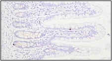
The human GLP-2R was localized to enteroendocrine cells in the human gastrointestinal tract.These findings imply that many of the actions of GLP-2 may be indirect, perhaps via GLP-2-stimulated liberation of multiple mediators of GLP-2 action from distinct gut endocrine cell populations.
GLP-2R+ gut endocrine cells in the human SB. To review the original data, see Enteroendocrine localization of GLP-2 receptor expression in humans and rodents. Gastroenterology. 2000 Sep;119(3):744-55
The enteroendocrine localization of GLP-2R expression described for human intestine has not been consistently detected in the rat or mouse gastrointestinal tract. Nevertheless, using two completely different antisera, Guan and colleagues localized GLP-2R expression to enteroendocrine cells and enteric neurons in the porcine and human intestine. Consistent with these anatomical findings, GLP-2 infusion stimulated intestinal blood flow and upregulated intestinal eNOS mRNA, protein, and phosphorylation. These investigators did not detect GLP-2R expression in subepithelial myofibroblasts-see GLP-2 Receptor Localizes to Enteric Neurons and Endocrine Cells Expressing Vasoactive Peptides and Mediates Increased Blood Flow. Gastroenterology. 2006 Jan;130 (1):150-164. Similarly, Ney and colleagues localized GLP-2R expressiob by immunohistochemistry to vagal afferents in the nodose ganglia, enteroendocrine cells, enteric neurons, and nerve fibers in the myenteric plexus as described in Localization and activation of glucagon-like peptide-2 receptors on vagal afferents in the rat. Endocrinology. 2007 May;148(5):1954-62
In rodents, the GLP-2R localizes to the enteric nervous system, or subepithelial myofibroblasts, but not the epithelium. Hence, although the majority of GLP-2 actions are also likely mediated via indirect mechanisms, the enteric nervous system, and not the gut endocrine system, appears to transduce the initial GLP-2 signal in rodents. To review the ENS localization data in mice, see Modulation of specific intestinal epithelial progenitors by enteric neurons. Proc Natl Acad Sci U S A. 2001 Oct 23;98(22):12497-502.
GLP-2 receptor expression has also been studied using a polyclonal antisera raised against the N-terminal region of the rat GLP-2R to identify GLP-2R+ cell types in the rat. Administration of 200 ug of human GLP-2 activated c-fos expression in the NTS and perivagal application of capsaicin eliminated the GLP-2-induced fos expression. In contrast, capsaicin treatment did not attenuate the trophic effects of exogenous GLP-2 on the rat gastrointestinal epithelium in the setting of TPN-associated mucosal atrophy. GLP-2R expression was documented in nerve cells within the nodose ganglion, enteric neurons and enteroendocrine cells of the rat gastrointestinal mucosa. See Localization and activation of GLP-2 receptors on vagal afferents in the rat. Endocrinology. 2007 Jan 18; [Epub ahead of print]
Catherine Orskov and colleagues used two different polyclonal antisera directed at different regions of the GLP-2 receptor to study the localization of GLP-2R in the small and large bowel from several different species, including rats, mice, marmoset and humans. The GLP-2R was localized predominantly to subepithelial myofibroblasts, with only weak staining detectable in enteric neurons. A subset of GLP-2R+ cells costained with antisera against KGF and co-administration of immunoneutralizing antisera against KGF together with GLP-2 abolished the trophic effects of GLP-2 in the colon but not the small bowel. See GLP-2 stimulates colonic growth via KGF, released by subepithelial myofibroblasts with GLP-2 receptors. Regul Pept. 2005 Jan 15;124(1-3):105-12
Bernardo Yusta and colleagues from the Drucker lab present further insights into GLP-2 receptor activation using cells transfected with the rat GLP-2 receptor. Identification of glucagon-like peptide-2 (GLP-2)-activated signaling pathways in baby hamster kidney fibroblasts expressing the rat GLP-2 receptor. J Biol Chem. 1999 Oct 22;274(43):30459-67. These studies demonstrate that at high doses, GLP-2 induces cell proliferation and immediate early gene expression, but these same effects were markedly attenuated at much lower doses in the 1 nM range. Evidence for GLP-2 activation of both PKA- and AP-1-dependent pathways is also presented.
Multiple studies have been directed at understanding the role of GLP-2 in the control of apoptosis. Induction of apoptosis in BHK-GLP-2R cells using cycloheximide is prevented by pretreatment of the cells with GLP-2. Furthermore, GLP-2 inhibits the activation of caspase-8 and caspase-9 activity, and markedly attenuates cytochrome C release, caspase-3 cleavage, and cytochrome c liberation into the cytoplasm. The net result of these effects is a GLP-2-mediated reduction in cell mortality in vitro. These findings suggest that GLP-2 exerts both direct and indirect anti-apoptotic actions in vivo. For the details of the specific experiments, see Yusta, B., Boushey, R.P., Drucker, D.J. The Glucagon-like peptide-2 receptor mediates direct inhibition of cellular apoptosis via a PKA-independent pathway J. Biol. Chem. Nov 2000 275: 35345-35352 and GLP-2 receptor activation engages Bad and glycogen synthase kinase 3 in a protein kinase A-dependent manner and prevents apoptosis following inhibition of phosphatidylinositol 3-kinase. J Biol Chem. 2002
There
is very little information available about mechanisms controlling GLP-2R
desensitization and resensitization. Jen Estall studied GLP-2R
desensitization in BHK fibroblasts and DLD-1 colon cancer cells transfected with
the rat and human GLP-2Rs. The GLP-2R
appears to exhibit unique properties and traffics via a clathrin- and 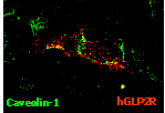 dynamin-independent, lipid raft-dependent pathway, as described in Lipid
Raft-dependent Glucagon-like Peptide-2 Receptor Trafficking Occurs Independently
of Agonist-induced Desensitization. Mol Biol Cell. 2004
Aug; 15(8):3673-87
dynamin-independent, lipid raft-dependent pathway, as described in Lipid
Raft-dependent Glucagon-like Peptide-2 Receptor Trafficking Occurs Independently
of Agonist-induced Desensitization. Mol Biol Cell. 2004
Aug; 15(8):3673-87
The GLP-2 Receptor carboxyterminus plays a key role in the intracellular trafficking of the receptor, but is not required for GLP-2R desensitization, despite engagement of beta arrestin-2, as described in The GLP-2R C-terminus modulates beta-arrestin-2 association, but is dispensable for ligand-induced desensitization, endocytosis and G-protein-dependent effector activation. J Biol Chem. 2005 Apr 6; [Epub ahead of print]
Although cyclic AMP formation is a hallmark of the cellular response to GLP-2, in some cells, the GLP-2R is also coupled to MAP kinase activation through distinct signaling pathways. The GLP-2R-induced activation of ERK1/2 was not mediated through Galphas, adenylyl cyclase, or transactivation of the epidermal growth factor receptor, but was pertussis toxin sensitive, inhibited by dominant negative Ras, and dependent on betagamma-subunits as outlined in The HeLa cell glucagon-like peptide-2 receptor is coupled to regulation of apoptosis and ERK1/2 activation through divergent signaling pathways. Mol Endocrinol. 2005 Feb;19(2):459-73
The factors that regulate control of GLP-2 receptor expression in the GI tract are poorly understood. Exogenous administration of GLP-2 was associated with a 3-fold increase in levels of GLP-2 R mRNA transcripts in the ileum of rats following majro small bowel resection and GLP-2R expression was localized to VIP+ and eNOS+ enteric neurons and serotonin+ gut endocrine cells. See Exogenous Glucagon-Like Peptide-2 (GLP-2) Augments GLP-2 Receptor mRNA and Maintains Proglucagon mRNA Levels in Resected Rats JPEN J Parenter Enteral Nutr. 2008 May-Jun;32(3):254-65
Several studies have demonstrated direct effects of GLP-2 on intestinal epithelial cell lines, however the cloned GLP-2 receptor has not been demonstrated to be expressed in the cells under study. For example, see Signaling mechanisms of glucagon-like peptide 2-induced intestinal epithelial cell proliferation. J Surg Res. 2000 May 1;90(1):13-8 and Glucagon-like peptide 2 stimulates intestinal epithelial proliferation in vitro. Dig Dis Sci. 2002 May;47(5):1135-40.
How does GLP-2 activate cell growth in the gut mucosa? Studies employing rat intestinal mucosal cells exhibited a cAMP response and a modest proliferative response following exposure to GLP-2, however no changes in p44/p42 MAPK phosphorylation or the levels of cytosolic calcium were detected in the mucosal cell preparation. Whether the GLP-2 receptor is normally expressed in these cells, or induced by the dissociation and cell culture conditions, remains unclear. See Glucagon-like peptide-2 receptor activation in the rat intestinal mucosa. Endocrinology. 2003 Oct;144 (10): 4385-92.
A role for VIP as a downstream mediator of GLP-2 actions in enteric neurons has been identified by Sigalet, Sharkey and colleagues. Exogenous GLP-2 ameliorates inflammation in rodents through VIP-dependent pathways independent of changes in cell proliferation Enteric neural pathways mediate the anti-inflammatory actions of glucagon-like peptide 2 Am J Physiol Gastrointest Liver Physiol. 2007 Jul;293(1):G211-21.
de Heuvel and colleagues subsequently showed that GLP-2 enhanced VIP expression in cultures of enteric neurons from the rat and mouse colon (submucosal plexus) and GLP-2R expression was identified by immunohistochemistry on populations of enteric neurons and glial cells. GLP-2 treatment rapidly increased the phosphorylation of AKT, Erk1/2 and p70S6K, but did not change levels of cyclic AMP in the cultures. Inhibition or genetic deletion of PI3kg eliminated the effects of GLP-2 on VIP expression. GLP-2 also rapidly induced growth factor-related gene expression (Igf1r, EGFR, Erbb, IGF-1, amphiregulin, epiregulin, EGF, hbEGF, betacellulin, and neuroregulins. Glucagon-like Peptide 2 induces vasoactive intestinal polypeptide expression in enteric neurons via phophatidylinositol-3 kinase-γ signalling Am J Physiol Endocrinol Metab. 2012 Aug 14.
Genetic variation in the GLP2R has been linked to level of hemoglobin F in humans with sickle cell disease Genome-wide association study identifies genetic variants influencing F-cell levels in sickle-cell patients Am J Human Genetics 2011 Apr;56(4):316-2 and GLP2R was identified as a potential locus contributing risk to development of type 2 diabetes in human European subjects An Expanded Genome-Wide Association Study of Type 2 Diabetes in Europeans Diabetes. 2017 May 31. pii: db161253 The mechanisms linking GLP2R biology to these phenotypes remain unknown